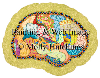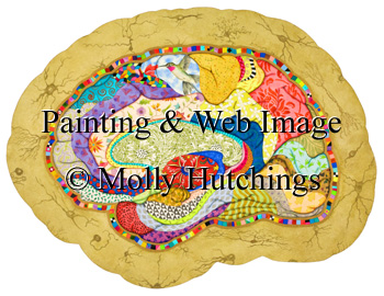
Quilter's Brain I and IIThe watercolor diptych, Quilter's Brain I & II, was commissioned by a Los Angeles neuroradiologist in 2007. The design inspiration was to represent a human brain using distinctive quilt patterns, with boundaries based on the Brodmann areas* of the brain cortex. 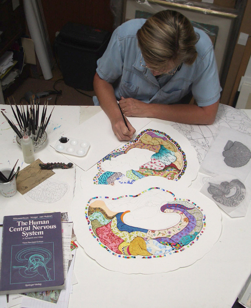
I started by making accurate drawings of the brain shapes in pencil, and then painted my designs with watercolors so that the patterns appear to wrap around and through the sulci. I wanted the painting to be as anatomically correct as possible, so I carefully indicated where sulci and Brodmann boundaries intersect. The diptych consists of two views of a brain: a lateral view of the outside, as seen from the side, and a medial view of the inside half, also viewed from the side. The lateral view includes a pattern with retinal neurons in one of the optical areas of the brain, while the outside border of the medial view features a variety of neuron types. The rest of the patterns are fabric-like and include creatures and colors from the quilter's brain. * Korbinian Brodmann first defined the Brodmann areas in 1909, based on the organization of the brain cortex when stained for nerve cells.
|